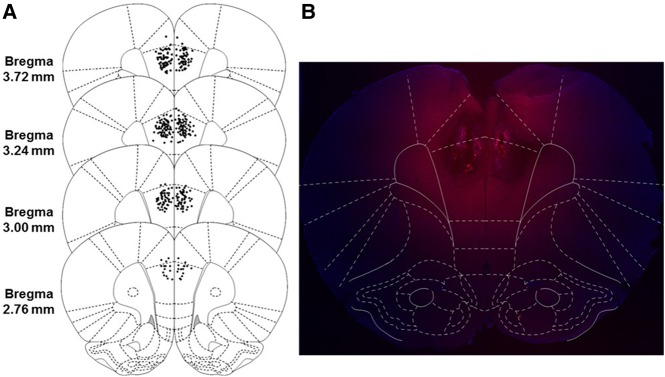
Figure 1.
Schematic representation of the majority of injection cannula tip placements in the mPFC for Experiments 1–4 (left, A) with visualization of the drug spread using the fluorescent muscimol BODIPY-TMRX (right, B). (A) Animals included in final analyses are represented by filled black dots. Placements ranged from 4.20 to 2.52 mm from bregma, with cannula placements from 10 animals being excluded for misses either anterior or posterior to this range (not shown). (B) Image taken from an animal infused with fluorescent muscimol into the mPFC, overlaid with a digital mPFC plate to examine the dorsal–ventral and medial–lateral drug spread. The anterior–posterior spread (not shown) was analyzed in sagittal sections and ranges from 1.75 to 2 mm from the cannula tract in any direction. (Reprinted from Paxinos and Watson 2007 with permission from Elsevier © 2007.)

