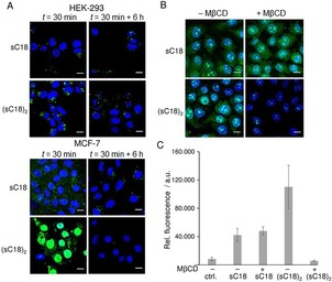
Figure 3.

A) CLSM images of MCF‐7 and HEK‐293 cells after 30 min with 10 μm CF‐sC18 or CF‐(sC18)2, and after 6 h in peptide‐free medium. Blue: Hoechst nuclear stain, green: CF‐labeled peptides. Scale bar: 10 μm. B) CLSM images of MCF‐7 incubated with 5 μm CF‐sC18 or 1 μm CF‐(sC18)2 for 30 min after cholesterol depletion using 10 mm methyl‐β‐cyclodextrin (MβCD) for 1 h. Blue: Hoechst nuclear stain, green: CF‐labeled peptides. Scale bar: 10 μm. C) Flow cytometric analysis of cellular uptake of CF‐labeled peptides sC18 and (sC18)2 in MCF‐7 cells incubated for 30 min at 37 °C with or without cholesterol depletion. Experiments were performed in triplicate with n=3.
