Figure 1.

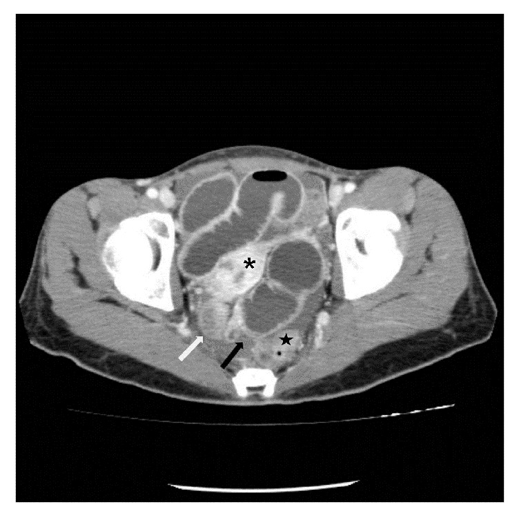
Axial CT image shows distended proximal bowel loop and collapsed distal loop (white arrow) at a transition zone (black arrow) between the uterus (asterisk) and the rectum (star).

Axial CT image shows distended proximal bowel loop and collapsed distal loop (white arrow) at a transition zone (black arrow) between the uterus (asterisk) and the rectum (star).