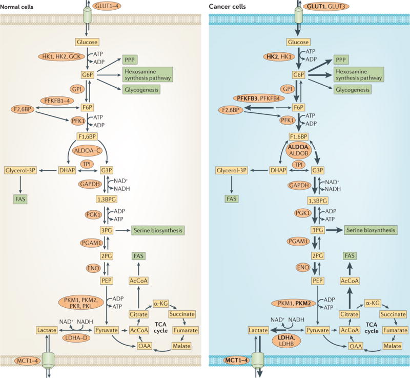
Figure 1. Changes that occur in glucose metabolism of cancer cells.

Compared with normal cells (left), the flux of glucose metabolism and glycolysis is accelerated in cancer cells (right) by preferential expression of transporters and enzyme isoforms that drive glucose flux forward and to adapt to the anabolic demands of cancer cells. Enzymes that catalyse the metabolic reactions are shown in ovals. Enzymes that are predominant in cancer cells are shown in bold. The thickness of the arrows indicates relative flux. 1,3BPG, 1,3-bisphosphoglycerate; 2PG, 2-phosphoglycerate; 3PG, 3-phosphoglycerate; α-KG, α-ketoglutarate; AcCoA, acetyl-CoA; ALDO, aldolase; DHAP, dihydroxyacetone-phosphate; ENO, enolase; F1,6BP, fructose-1,6-bisphosphate; F2,6BP, fructose-2,6-bisphosphate; F6P, fructose-6-phosphate; FAS, fatty acid synthesis; G3P, glyceraldehyde-3-phosphate; G6P, glucose-6-phosphate; HK, hexokinase; LDH, lactate dehydrogenase; GAPDH, glyceraldehyde-3-phosphate dehydrogenase; GCK, glucokinase; GLUT, glucose transporter; glycerol-3P, glycerol-3-phosphate; GPI, glucose-6-phosphate isomerase; MCT, monocarboxylate transporter; OAA, oxaloacetate; PEP, phosphoenolpyruvate; PFK1, phosphofructokinase 1; PFKFB, 6-phosphofructo 2-kinase/fructose-2,6-bisphosphatase; PGAM1, phosphoglycerate mutase 1; PGK1, phosphoglycerate kinase 1; PK, pyruvate kinase; PPP, pentose phosphate pathway; TCA, tricarboxylic acid; TPI, triosephosphate isomerase.
