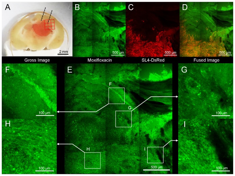
Fig. 5.
Moxifloxacin-based TPM image of mouse brain that contains metastatic tumor. (A) SL4-DsRed tumor was mainly located ln left cerebral hemisphere. Red 3 × 3 rectangular region was imaged with TPM. (B) TPM image after topically treated moxifloxacin (green). (C) TPM Red fluorescence image confirming by Ds-Red labeled cancer cells. (D) Merged fluorescence image with moxifloxacin and SL4-DsRed. (E) Four regions of interest (ROIs) are magnified: (F) Multiple cellular bodies were detected in hippocampus. (G) Round, condensed cellular structure was shown. (H) Tumor lesion showed high cellular density. (I) Border of tumor and normal tissue. The cellular density of tumor area was significantly higher than adjacent normal tissue.

