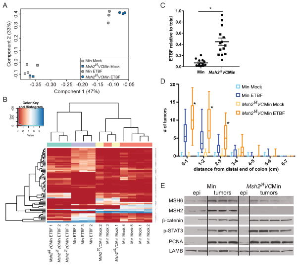
Figure 4. Inflammation-induced tumorigenesis is increased in the distal colon of mice with altered Msh2 expression.
A) PCA analysis of 16S microbiome sequencing of DNA from stool samples from WT/Min or Msh2l/lVC/Min mice 8 weeks post mock or ETBF. B) Heatmap representing the unsupervised hierarchical clustering of 65 OTUs found to be differentially abundant by one pair-wise comparison (rows) in individual stool samples (columns) from indicated mice treated as in A. C) ETBF abundance in stool relative to total bacterial DNA by qPCR. Symbols represent data from individual mice. Horizontal line is mean +/− SEM. N≥13. *p<0.05. D) Tukey box plots of tumor counts by cm in WT/Min or Msh2l/lVC/Min mice 8 weeks after mock or ETBF. N≥8. *p<0.05. E) Whole cell protein lysate from distal colon epithelium or tumors from mice of the indicated genotypes 8 weeks post-mock or ETBF inoculation, respectively, were blotted for the indicated proteins. See also Figure S4.

