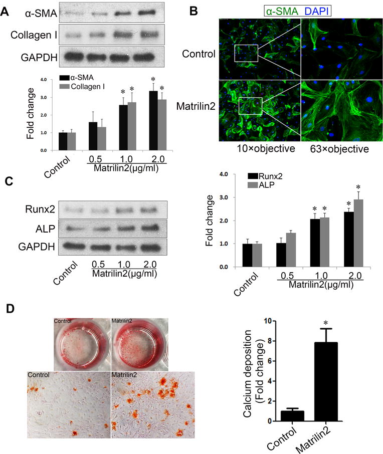
Figure 2. Matrilin2 induces myofibroblastic transition and upregulates pro-osteogenic activity in valvular interstitial cells (VICs).

A. VICs from normal human aortic valves were treated with recombinant matrilin2 (2.0 μg/ml) for 48 h. Matrilin2 induced myofibroblastic transition of VICs, as characterized by enhanced expression of α-SMA and collagen I. B. VICs had greater amounts of α-SMA-containing stress fibers (green) following matrilin2 stimulation. 4′, 6-diamidino-2-phenylindole (DAPI) was used for nucleus counterstaining (blue). C. Matrilin2 increased the expression of Runx2 and ALP in VICs in a dose-dependent manner. D. VICs were incubated with matrilin2 for 21 days in conditioning medium (containing 10 mmol/L β-glycerophosphate, 10 nmol/L dexamethasone, 4.0 μg/ml cholecalciferol and 8 mmol/L CaCl2) to examine calcium deposit formation. Images of Alizarin red S staining (10× objective for the high power images) and spectrophotometric analysis show that prolonged stimulation with matrilin2 increased calcium deposit formation. Mean ± SE; n=5 separate experiments using distinct cell isolates; *P<0.05 vs. control.
