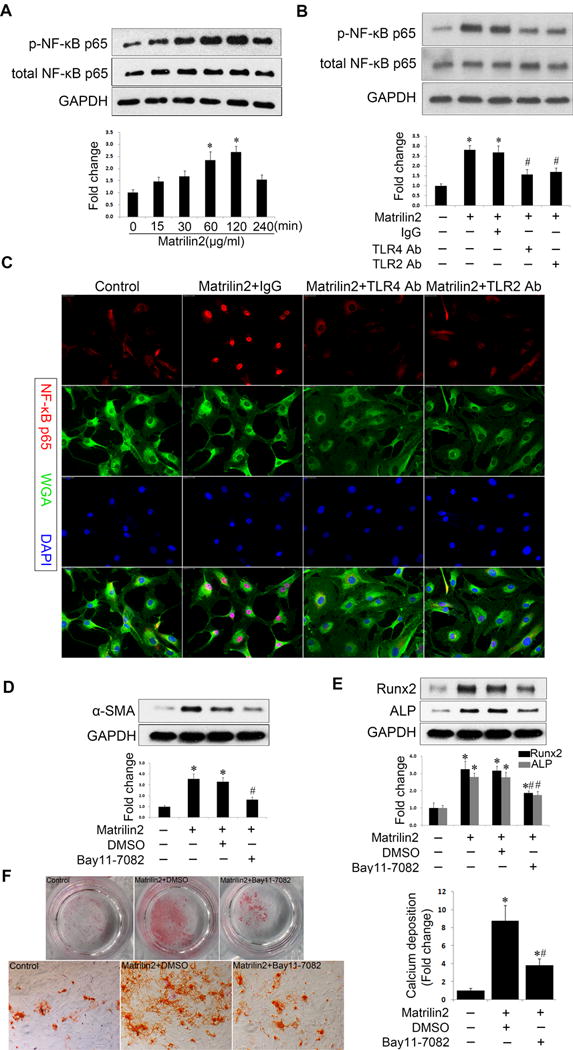
Figure 4. NF-κB plays an important role in matrilin2-induced valvular interstitial cell (VIC) phenotypic transition.

A. VICs from normal human aortic valves were treated with matrilin2 (2.0 μg/ml) for 15 min to 4 h to examine the time course of NF-κB phosphorylation. Phosphorylation of NF-κB p65 occurred mainly at 1 and 2 h following matrilin2 treatment. B and C. VICs were treated with matrilin2 (2.0 μg/ml) for 2 h in the presence or absence of neutralizing antibodies against TLR4 (10 μg/ml) and TLR2 (10 μg/ml). Immunoblots show that neutralizing antibodies against TLR4 and TLR2 suppressed NF-κB p65 phosphorylation following matrilin2 stimulation. Representative images of immunofluorescence staining of NF-κB p65 (red) show that NF-κB p65 intranuclear translocation induced by matrilin2 was reduced by neutralizing antibodies against TLR4 and TLR2. Alexa 488-tagged wheat germ agglutinin (WGA) was used to outline cells (green). 4′, 6-diamidino-2-phenylindole (DAPI) was used for nuclear counterstaining (blue). Original magnification, 40× objective. D and E. NF-κB inhibitor, Bay 11-7082 (10 μM), reduced the levels of α-SMA, Runx2 and ALP in cells exposed to matrilin2 (2.0 μg/ml) for 48 h. F. To examine calcium deposit formation, VICs in the conditioning medium were treated with matrilin2 (2.0 μg/ml) for 21 days in the presence or absence of Bay 11-7082 (10 μM). Alizarin red S staining shows that NF-κB inhibition reduced the formation of calcium deposits in cells treated with matrilin2 (10× objective for the high power image). Mean ± SE; n=4 separate experiments using distinct cell isolates; *P<0.05 vs. control; #P<0.05 vs. matrilin2 + non-immune IgG or matrilin2 + DMSO.
