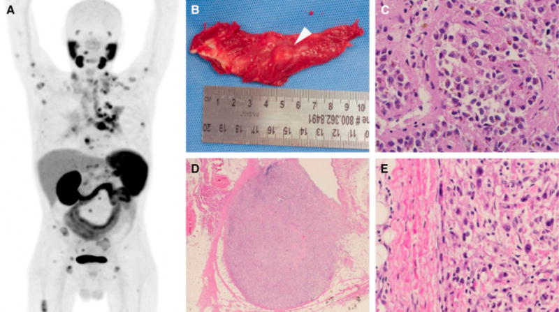
Fig. 1.

(a) Maximal intensity projection of the imaged patient. Normal biodistribution of the radiotracer includes the salivary glands, lacrimal glands, liver, spleen, kidneys, and small bowel. Excreted radiotracer is also observed in the ureters and bladder. All other sites of radiotracer uptake represent putative sites of metastatic clear cell renal cell carcinoma (ccRCC). (b) Gross image of a 10-mm nodule of ccRCC found within the right triceps muscle. This lesion was occult on conventional imaging and (c) histologically confirmed to be ccRCC (×200). (d) Low-power (×40) and (e) high-power (×200) images of a 6-mm intramammary lymph node with metastatic ccRCC observed only on 18F-DCFPyL positron emission tomography/computed tomography.
