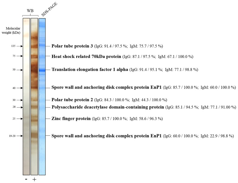
Fig 2. Example of negative and positive Western blot (WB) patterns for extracted Encephalitozoon cuniculi proteins using goat anti-rabbit IgG conjugate.
The identification of the proteins in the SDS-PAGE gel (right panel) corresponding to the bands of interest on the WB (left panel) are listed on the right. In brackets: the respective sensitivity and specificity for IgG and IgM detection for each WB band. The protein identified as spore wall and anchoring disk complex protein EnP1 at 17–19 kDa was actually a fragment of the one found in 40 kDa. Abbreviations: -, Negative; +, Positive; kDa, Kilodalton; WB, Western blot.

