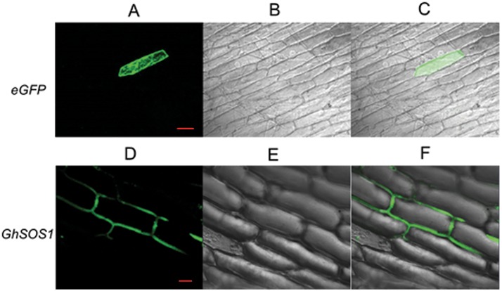
Fig 3. Localization of GhSOS1 in onion epidermal cells.
A-C: Onion epidermal cells transformed with 35S::GFP. Bar: 100 μm. D-F: Onion epidermal cells transformed with 35S::GFP-GhSOS1. Bar: 50 μm. A and D: Dark field images for the detection of GFP fluorescence. B and E: Light field microscopy images to display morphology. C and F: Superimposed light and dark field images.

