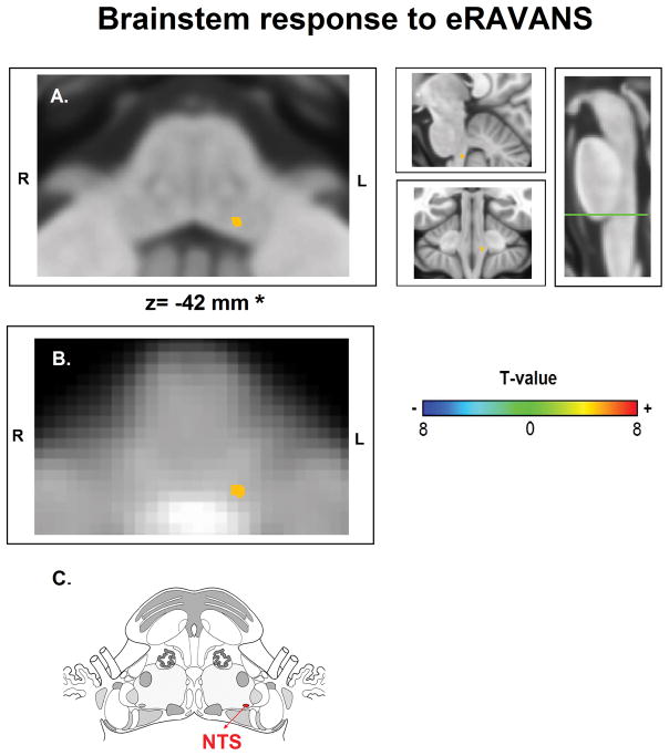
Figure 4.
FMRI brainstem response to Respiratory-gated Auricular Vagal Afferent Nerve Stimulation (RAVANS). Activation found in the left nucleus tractus solitarius (NTS) when contrasting RAVANS vs SHAM stimulation overlayed on (A) MNI-space template and (B) group-averaged functional images rotated for consistency with (C) the Duvernoy brainstem atlas. Right panel of (A) shows the level of the axial slice in green (Obex+18 mm) and sagittal and coronal views of NTS activation. *z-coordinates refer to MNI space results and not the particular image visualized.

