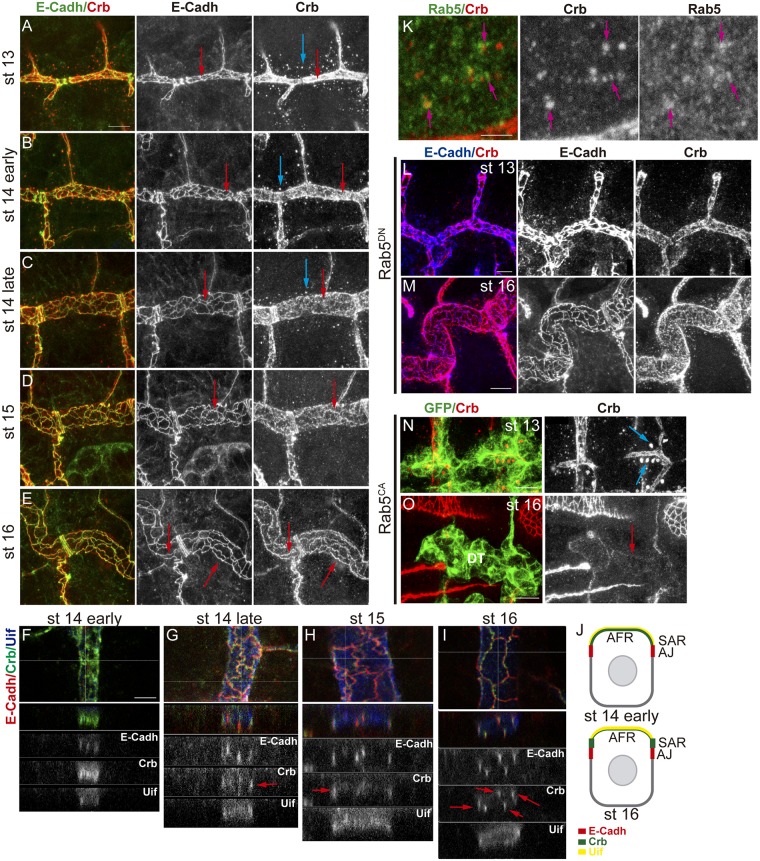
Fig 4. Subcellular accumulation of Crb during tracheal development.
(A-E) Lateral views of wild type embryos at indicated stages stained for E-Cadh (green, white) and Crb (red, white), showing 1–2 tracheal metameres. At early stages, while E-Cadh is already detectable in the junctional area (red arrows), Crb mostly accumulates in the AFR. Crb accumulation becomes enriched in the SAR as development proceeds (E). At early stages, abundant vesicles containing Crb are detected (blue arrows), and they decrease as development proceeds. Scale bar 7,5 μm (F-I) Z-reconstructions of the DT of embryos at the indicated stages stained for E-Cadh (red, white), Crb (green, white) and Uif (blue, white). Horizontal line in the upper panel indicates the position of the Z- reconstruction. E-Cadh always localise to the AJs, in the apicolateral membrane, visualised as lines spanning the AJs. Uif localises in the most apical membrane. Note the evolution of Crb pattern that becomes more enriched in the SAR (red arrows, partially colocalising with E-Cadh) as development proceeds. Scale bar 5 μm (J) Diagram representing a Z-section of a DT cell at early and late stages. The apical region of the tracheal cells facing the lumen (AFR) accumulates Uif (yellow). E-Cadh accumulates in AJs (red). Crb (green) first accumulates more in the Uif region and later becomes enriched in the SAR. (K) Wild type embryo stained with Rab5 (green, white) and Crb (red, white) antibodies. Many Crb vesicles co-stain with Rab5 (pink arrows). Scale bar 2,5 μm (L,M) Lateral views of embryos with downregulated Rab5 activity at the indicated stages stained for E-Cadh (blue, white) and Crb (red, white) showing 1–2 tracheal metameres. At early stages Crb vesicles are absent (G), and at late stages (H) Crb is not enriched in the SAR. Scale bar G 5 μm, H 7,5 μm (N,O) Lateral views of embryos with activated Rab5 activity at the indicated stages stained for GFP (green) and Crb (red, white) showing 2 tracheal metameres. At early stages huge Crb vesicles are detected (blue arrows in I), but at late stages (J) Crb is almost absent mainly from the DT region. Scale bar I 7,5 μm, J 10 μm.

