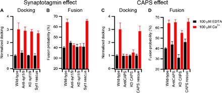
Fig. 2. Molecular origin of DCV calcium response.

The role of synaptotagmin (A and B) and CAPS (C and D) on DCV docking and fusion in the presence of 100 μM EDTA (black) or 100 μM Ca2+ (red). Wild-type preparations of DCVs were compared either to those that were treated with function-blocking antibodies or to those that were purified from cells subjected to shRNA-mediated knockdown (KD). In the latter case, expression of RNAi-resistant syt1 or CAPS-1 was used as a control. Black and red bar data were obtained in the absence and presence of Ca2+, respectively. Table S7 contains a summary of events, and table S8 contains fitting results. As indicated in Materials and Methods, docking values for preparations of DCVs from wild-type, knockdown, and RNAi rescue cell lines were individually normalized to the value obtained in the presence of 100 μM EDTA, enabling comparison of the relative effects elicited by calcium among the different preparations. This strategy does not enable us to rule out the possible effects of syt or CAPS knockdown on docking in the absence of calcium. For antibody-treated samples, we observed no significant effect on docking in the absence of calcium.
