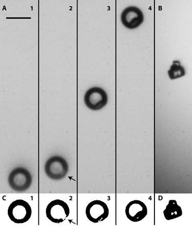
Fig. 4. Bright-field imaging of collision-induced contact efflorescence.

Crystal nucleation and growth of NaCl following contact with 400-nm (−)PSL CN at 57 ± 1% RH. Scale bar, 10 μm. (A) Frame 1 shows the aqueous droplet before contact. The droplet was deliberately positioned at the bottom of the frame to capture the crystal growth process as the droplet moved upward because of loss of water mass. Frame 2 shows the droplet ~20 ms after contact. Crystal growth is apparent near the droplet surface (where indicated by the arrow). Frames 3 and 4, ~90 and 180 ms after contact, respectively, show the progressive growth process. At this RH, complete NaCl efflorescence occurs at ~1 s. This particular particle did not stay trapped after contact efflorescence. (B) A fully effloresced NaCl crystal that remained trapped after contact efflorescence. (C and D) Binary contrast images of the particle shown in each frame of (A) and (B).
