
Fig. 2.
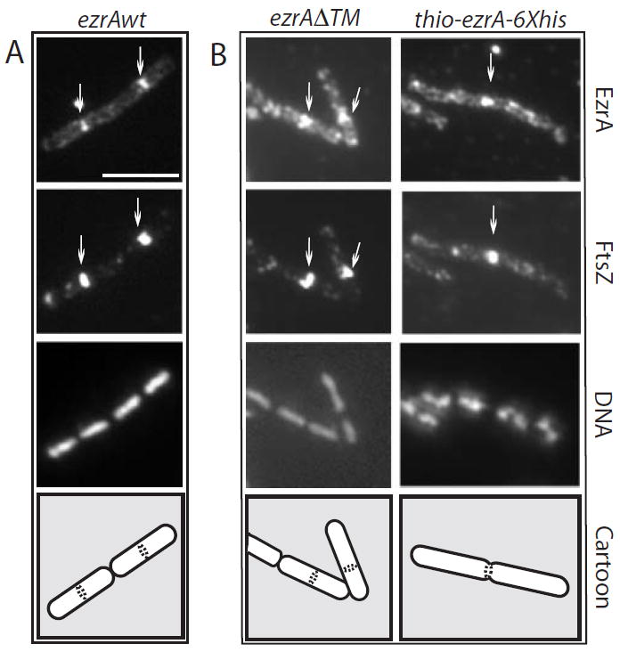
The transmembrane anchor is dispensable for EzrA localization. Cells were grown to mid-exponential phase (OD600 = ≈ 0.5) in the presence of IPTG and fixed and stained for immunofluorescence microscopy using antiserum against FtsZ (Levin and Losick, 1996) or EzrA (this work). A single field of view is shown in each column. From top to bottom: EzrA, FtsZ, DNA and a schematic showing cell boundaries and the position of the EzrA/FtsZ rings.
A. ezrA∷Pspachy-ezrA (PL923).
B. ezrA∷Pspachy-ezrAΔTM (PL925). The high background in the anti-EzrA panels in (B) results from cytoplasmic staining of EzrA in the absence of the transmembrane domain. Scale bar = 5 μM.