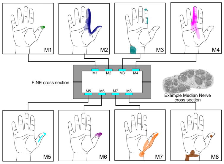
Figure 3.
The cross section of the FINE implanted on the median nerve of subject 2 and the corresponding, channel-specific percept areas are shown. The example nerve cross section shown is from a comparable location taken from human histology studies [31]; it is not from subject 2. Multiple measures, including suprathreshold responses up to week 56, are shown for each channel. Most channels produce sensation in stable and characteristic percept areas, suggesting that fascicles at the level of the implant retain somatotopic organization. A similar drawing is not available for subject 1 because the exact order of channels in his cuff electrodes is unknown.

