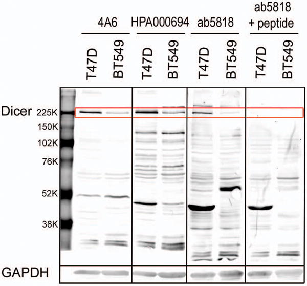
Figure 2. Antibody comparison of Dicer expression in ER+ versus triple-negative breast cancer cell lines.

All blots were scanned at similar intensity setting. Whole cell lysate of T47D (ER+) and BT549 (triple-negative) breast cancer cell lines probed for Dicer with 4A6, HPA000694, ab5818, and ab5818+10× blocking peptide (Dicer 217kD band highlighted in red box). GAPDH loading control corresponding to the above membrane 35 kD.
