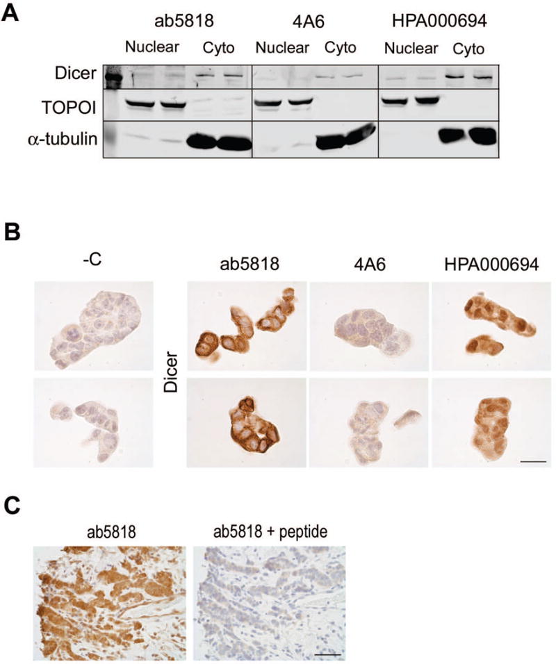
Figure 4. Antibody detection of Dicer in nuclear and cytoplasmic fractions and by IHC.

A) T47D cell extracts showing Dicer expression in the nuclear versus cytoplasmic fraction with all three antibodies. TOPOI used as a control for nuclear protein. α-tubulin used as a control for cytoplasmic protein. B) T47D cells, FFPE and stained for Dicer with the three antibodies. Bar = 25μm. C) ER+ Breast cancer stained for Dicer with ab5818 alone (left) and with ab5818+10× blocking peptide (right). Bar = 50μm.
