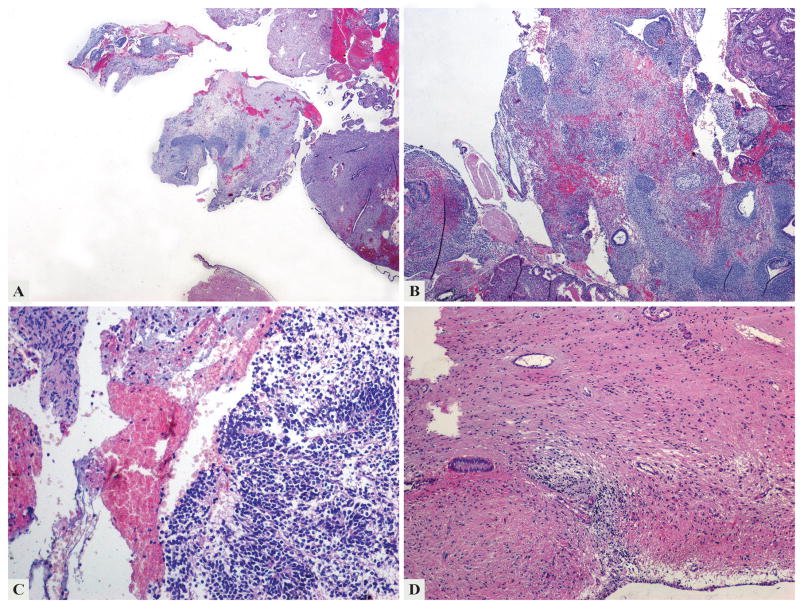
Figure 1.
The first specimen examined in 2003 contained multiple fragments of secretory endometrium and tumor tissue (a). The tumor was mainly composed of mature epithelial and mesenchymal tissue (b), but immature neuroectodermal components with poorly formed rosettes (c) and mature glial component surrounding endometrial glands (d) were also present.

