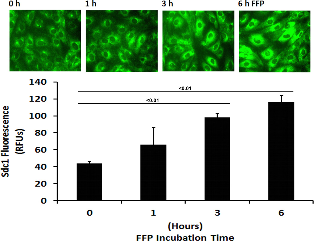
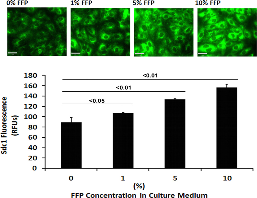
Fig. 1. FFP increases cell surface Sdc1 in a time- and dose-dependent manner.
Human lung microvascular endothelial cells were cultured in low serum medium (0.5% FBS) for 2 hours and then incubated with serum-free media containing the indicated percentages of FFP. Cells were stained with anti syndecan-1 antibody and images were captured with Nikon E800 fluorescence microscope. Original magnification, × 600. The relative fluorescence intensity was quantified using Quantity One (Bio-RAD) after converting the images to greyscales and reported as relative fluorescence units (RFU). A. Dose dependent increase in sdc1 and B. Time dependent increase in sdc1. Results are presented as means ± SEM, n=3/group.


