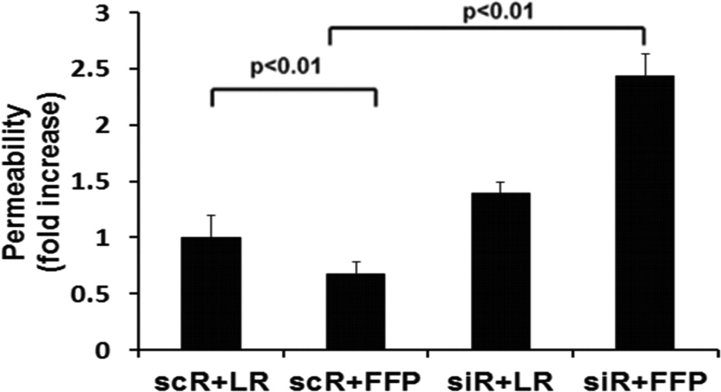
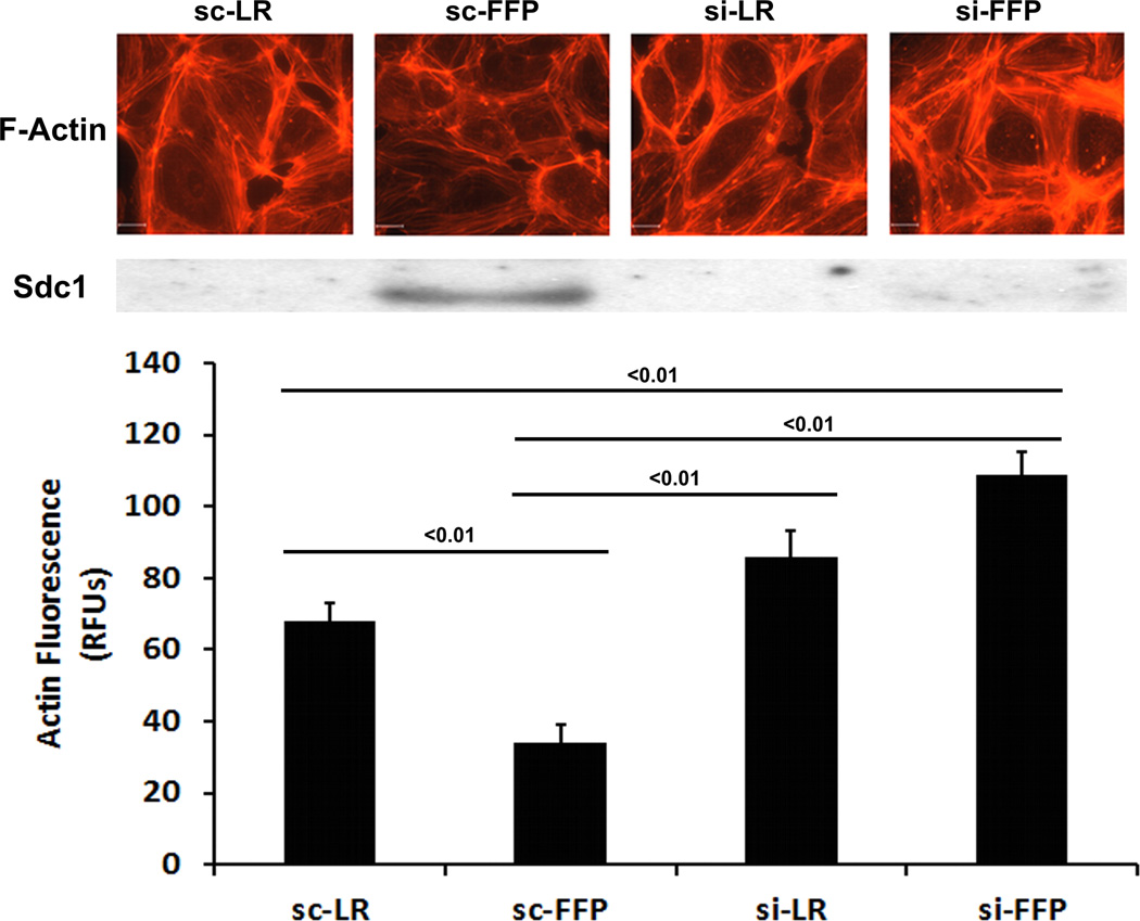
Fig. 2. FFP increases monolayer permeability and stress fiber formation in Sdc1-deficent endothelial cells.
Human lung microvascular endothelial cells were transfected with Sdc1 siRNA (siR) or scrambled RNA (scR) as control. A. In vitro permeability was assessed by fluoresceine-isothiocyanate cells treated with 10% LR or 10% FFP in serum free medium. B. Stress fiber formation, a marker of endothelial disruption, was examined by staining cells with Texas red-X phalloidin. Top panel: Images were captured with Nikon E800 fluorescence microscope. Original magnification, × 600. Middle panel: Sdc1 protein expression was assessed by Western blot. Bottom panel: the relative fluorescence intensity was quantified using Quantity One (Bio-RAD) after converting the images to greyscales and reported as relative fluorescence units (RFUs). Results are presented as means ± SEM, n=4/group.


