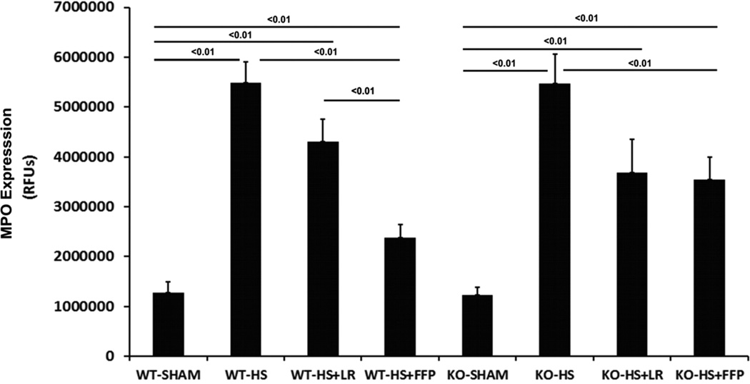
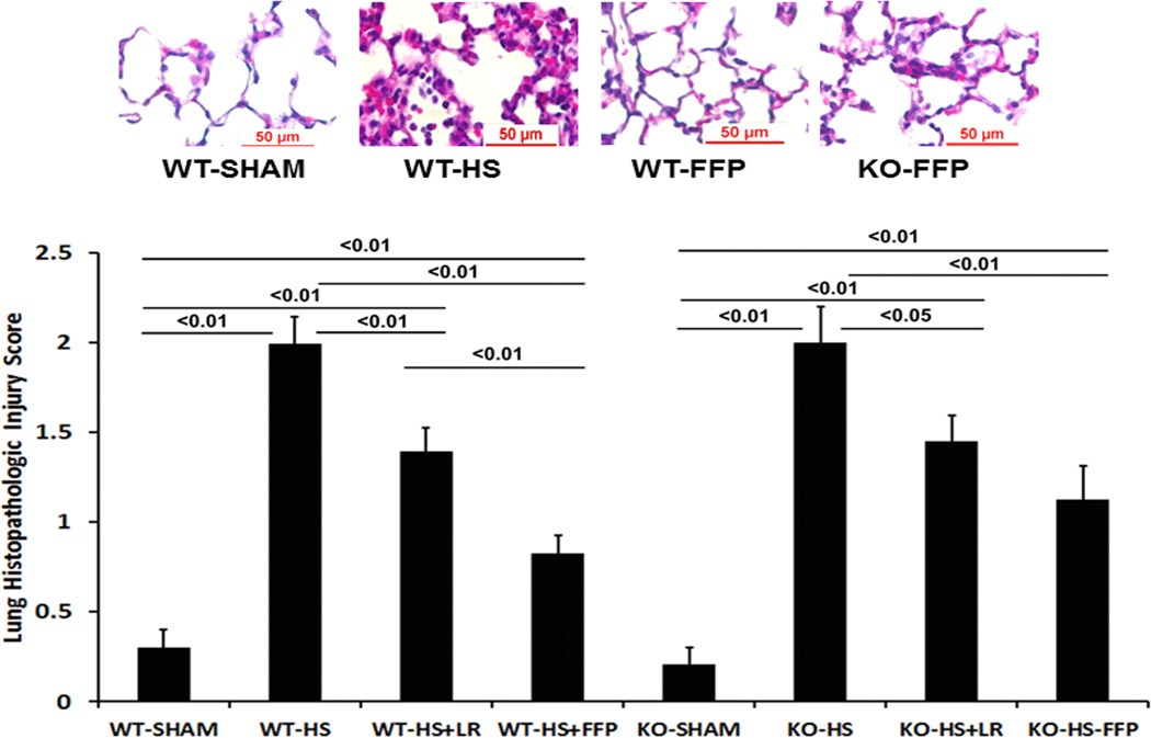
Fig. 4. Plasma mitigates lung inflammation and injury after hemorrhagic shock in wild-type but not syndecan-1 −/− mice.
Wild-type and syndecan-1 null mice underwent laparotomy and hemorrhagic shock followed by resuscitation with either LR or FFP. After 3 hours lungs were harvested for assessment of A. Myeloperoxidase (MPO) enzyme by immunofluorescent staining. Results are reported as relative fluorescence units (RFUs) or B. Lung histopathologic injury. Lungs sections were stained with hematoxylin and eosin and scored on alveolar thickness, capillary congestion, and cellularity. Representative images are shown in the upper panel and injury scores in the lower panel. Data are expressed as mean SEM, n=8 per group with significance indicated by lines over the respective groups. FFP, fresh frozen plasma; LR, lactated Ringer’s; WT, wild type; KO, Sdc1 knockout; HS, hemorrhage shock.


