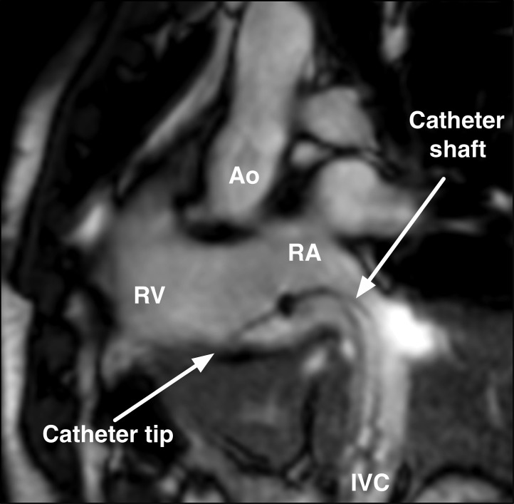
Figure 2: Passive Catheter Tracking.
An MR-compatible ablation catheter tip is visualised at the tricuspid valve annulus during a clinical ablation of the cavotricuspid isthmus for typical atrial flutter. The location of the catheter shaft is seen from IVC to RA. Ao = aorta; IVC = inferior vena cava; RA = right atrial; RV = right ventricular.

