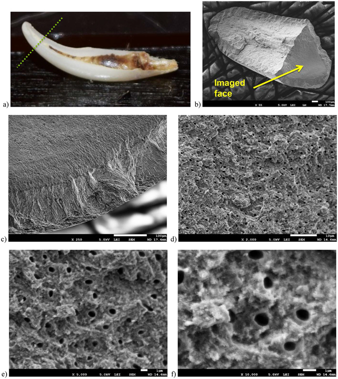
Figure 3.

Images of tooth sample after cracking open the first time and then after breaking tip and SEM micrographs of the interior surface of the tooth sample. (a) Optical image indicating where tip was broken. (b) SEM micrograph of broken tip and indication of imaged face for images (c–f). (c) Image of enamel – dentin interface at 250 X magnification. Scale bar is 100 μm. (d) Image of denting region at 2 KX magnification. Scale bar is 10 μm. (e) Image of dentin region at 5 KX magnification. Scale bar is 1 μm. (f) Image of dentin region at 10 KX magnification. Scale bar is 1 μm.
