
Figure 3.
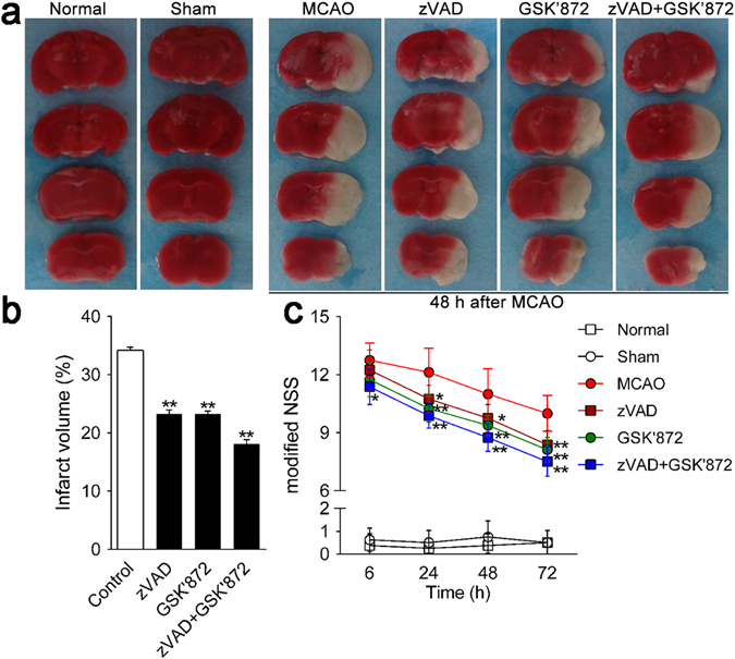
GSK’872 and combinative intervention attenuated ischemic brain injury and neurological deficits induced by MCAO in mice. (a) Ischemic areas were visualized by TTC staining at 48 h after MCAO (n = 3 per group). Normal tissue stained rose-pink, whereas infarct area appeared white. (b) Infarct volume was quantified by Image-Pro plus 6.0 software. (c) Neurological deficits were evaluated by mNSS at 6, 24, 48 and 72 h after MCAO (n = 9 per group). Data are presented as the mean ± SD. *p < 0.05, **p < 0.01 vs. control group.