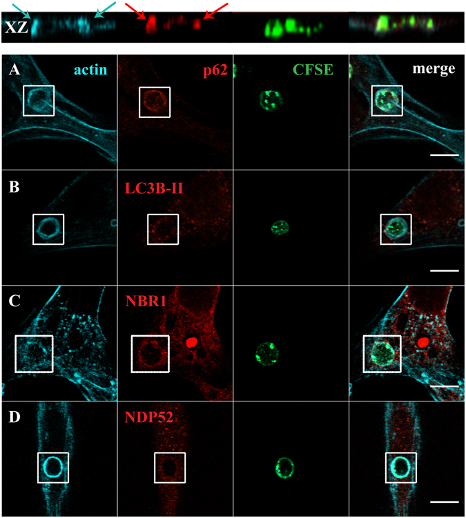
Figure 2.

Co-localization of LC3B-II and autophagy adaptor proteins with F-actin in phagosomes containing RBC. Non-professional phagocytes were fed with RBC for 30 min, fixed, and stained for F-actin with Phalloidin and for the endogenous LC3B or autophagy adaptors. (A–D) are representative images, obtained by confocal microscopy, of cells co-stained for F-actin and p62 (A), LC3B (B), NBR1 (C) or NDP52 (D). In A, side views (XZ) are merges of ten vertical sections of confocal stacks. Arrows indicate the nascent phagosome positive for F-actin (blue) and p62 (red). The first column represents cells stained for F-actin. The second column represents cells stained for the endogenous p62, LC3B-II, NBR1 or NDP52. The third column shows internalized RBC stained with CFSE. The fourth column represents merged images of F-actin with LC3B-II or autophagy adaptors and internalized RBC. The regions outlined by the boxes are nascent phagosomes. Bars, 10 µm.
