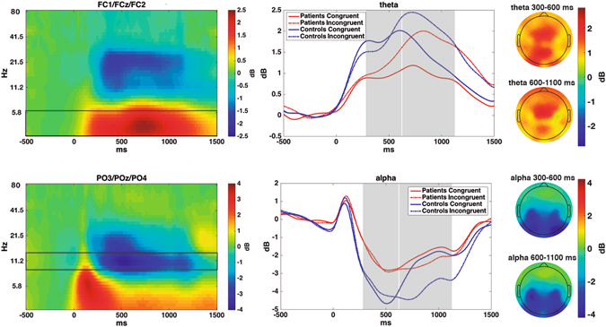
Figure 3.

Left: Time-frequency power modulations over midfrontal and posterior locations, averaged between groups and conditions. Boxes show the frequency ranges depicted in the left column. Middle: Time course of theta and alpha power modulations for each condition and group; Grey boxes show the time windows used for statistical comparisons. Right: Topographies -averaged between conditions and groups- for each frequency and time window.
