
Figure 1.
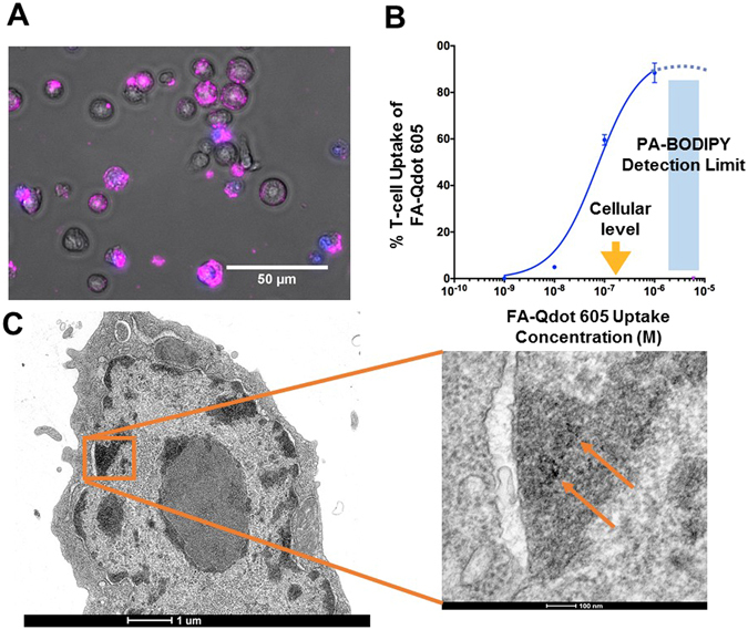
FA-Qdot 605 is taken up by activated T-cells. In (A), Epifluorescence imaging of T-cells in-vitro after incubation with FA-Qdot-605 conjugate (Purple), DAPI was added to solution to exclude cells undergoing apoptosis or those which have leaky membranes. In (B), Measurement of fatty acid Qdot-605 uptake at increasing concentrations as it compares to current FA-uptake detection via BODIPY dye. It is important to note that BODIPY uptake is less sensitive than cellular levels of palmitic acid (~200nM, yellow arrow). In (C), TEM image of FA-Qdot within a T-cell, image scale bars 1um and 100nm respectively, (4800x and 49000x zoom).