Figure 3.

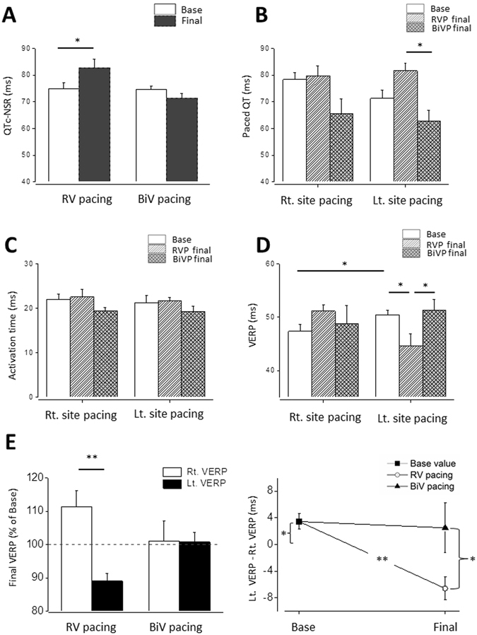
RV tachypacing prolongs native QTc interval and induces dispersion of VERP between the pacing sites in contrast to BiV tachypacing. Rats were implanted with RV pacing electrode and LV pacing electrode. Following a recovery period (7 d), rats were subjected to 72 h of either RV pacing or BiV pacing at a CL of 100 ms. (A) Measured QTc interval under sinus rhythm was evaluated in the conscious state using subcutaneous electrodes implanted on the back of the animals. Note prolongation of the QTc interval during RV tachypacing which did not occur in response to BiV pacing. Of note, statistical findings of QT and QTc measurements were similar (not shown), since the spontaneous heart rate of the rats did not change significantly by the pacing. (B) Paced QT interval following RV vs. BiV tachypacing. Following 72 h of pacing there was a significant difference between two ventricular pacing groups during Lt. site pacing. (C) Epicardial activation during Rt. site pacing and Lt. site pacing. Note absence of notable effects on this variable following 72 h of tachypacing. (D,E) VERP evaluated using double threshold stimulation intensity in the Rt. pacing site or the Lt. pacing site. Note that the baseline VERP is significantly longer in the Lt. pacing site. RV tachypacing caused a paradoxal effect by significantly reducing the Lt. VERP and an opposite tendency in the Rt. VERP creating a remarkable difference between the VERP of both sites. Note that such effect did not occur by similar application of BiV pacing.
