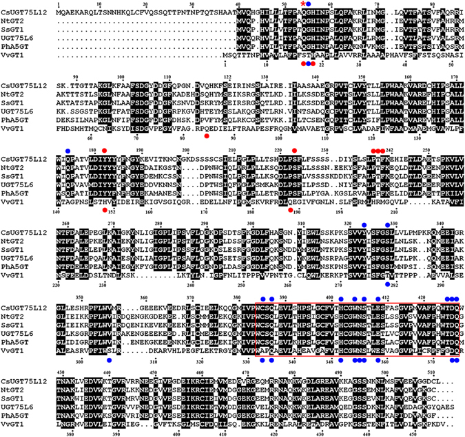
Figure 2.

Multiple alignment of deduced amino acid sequences of CsUGT75L12 with other UGTs. The UGTs' signature PSPG motifs were enclosed in a red box. Conserved residues between the CsUGT75L12 and others UGTs were indicated with a black column. The residue denoted with a red asterisk (*) was predicted to be a sugar donor and acceptor binding site for CsUGT75L12 above the aligned sequences. Conserved residues involved in sugar donor binding sites in CsUGT75L12 (above the aligned sequences) and VvGT1 (below the aligned sequences) were designated with blue dots. Conserved residues involved in sugar acceptor binding sites in CsUGT75L12 (above the aligned sequences) and VvGT1 (below the aligned sequences) were designated with red dots.
