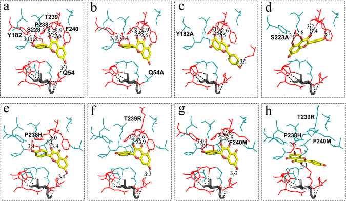
Figure 8.

Structure models of the sugar donor and acceptor binding sites of the CsUGT75L12 and the variants. Docking molecular interaction of the sugar donor molecule UDP-Glc (dark grey) and the sugar acceptor kaempferol (yellow). The amino acid residues involved in interaction with sugar donor and acceptor were highlighted in red, and the hydrogen bonds were indicated with black dotted lines. (a) CsUGT75L12 (WT); (b) single mutation (Q54A); (c) single mutation (Y182A); (d) single mutation (S223A); (e) single mutation (P238H); (f) single mutation (T239R); (g) single mutation (F240M); (h) triple mutation (P238H, T239R and F240M).
