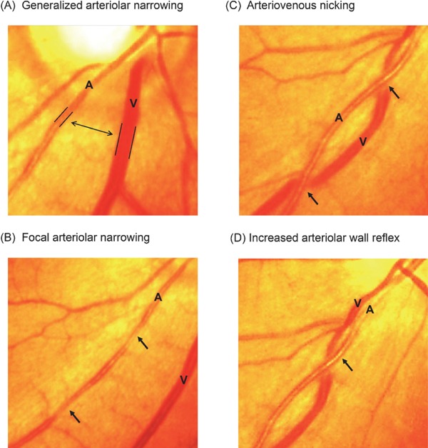
Fig. 2.

Example photographs of retinal vascular changes.
A, retinal arteriole; V, retinal venule; Example photographs of retinal vascular changes: (A) Generalized arteriolar narrowing: arteriolar-to-venular diameter ratio of 2:3 or lower; (B) Focal arteriolar narrowing: localized constrictions along the course of arterioles; (C) Arteriovenous nicking: narrowing of a venule as an arteriole crosses over it; and (D) Increased arteriolar wall reflex: an increased light reflex from the central portion of the retinal arteriolar surface; All photographs were modified from Iida and Kitamura (2009) with the authors' permission28).
