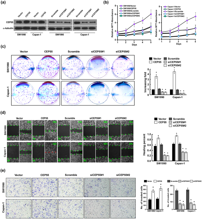
Figure 3.

CEP55 overexpression promoted pancreatic cancer (PANC) cells aggressiveness in vitro. (a) Expression of CEP55 protein as detected by Western blot analysis in SW1990 and Capan-1 cells upon transfection with CEP55 or CPE55 short interfering RNAs (siRNAs). GAPDH was used as the loading control. (b) MTT (3-(4,5-Dimethylthiazol-2-Yl)-2,5-Diphenyltetrazolium Bromide) assay of SW1990 and Capan-1 cell growth curves following CEP55 or CEP55 siRNAs transfection. (c) Representative micrographs (left panel) and quantification (right panel) of crystal violet-stained SW1990 and Capan-1 cell colonies after 14 day of colony formation. (d) Wound-healing assay (left panel) and quantification (right panel) of the wound distance to assess SW1990 and Capan-1 cell migration after transfection with CEP55 or CEP55 siRNAs. (e) Representative micrographs (left panel) and quantification of SW1990 and Capan-1 cell invasion in a Transwell matrix penetration assay. The quantification of the invaded cells is represented by the mean of three independent experiments. Bars represent the mean ± SD of three independent experiments. *P < 0.05.
