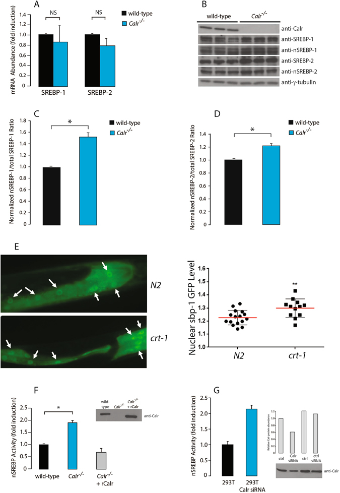
Figure 2.

SREBP expression and processing in the absence of calreticulin. (A) Q-PCR quantitative analysis of total SREBP-1 and SREBP-2 mRNA abundance in wild-type and Calr −/− cells. Results were normalized to 18S rRNA (internal control). NS, not significant (Student’s t-test). Representative of 5 biological replicates. (B) Immunoblot analysis of SREBP-1, nSREBP-1, SREBP-2 and nSREBP-2 protein in wild-type and Calr −/− cells. Anti-γ-tubulin antibodies were used as a loading control. Representative of 3 biological replicates. (C,D) Quantitative analysis of immunoblots showing the ratio of nuclear to total SREBP-1 (C) and total SREBP-2 (D) in wild-type and Calr −/− cells. The value for the total is the sum of precursor and nuclear forms of SREBP. *Indicates statistically significant differences: SREBP-1, p-value = 0.0017 (Student’s t-test); SREBP-2, p-value = 0.0218 (Student’s t-test). Representative of 3 biological replicates. (E) GFP:SBP-1 accumulation in the intestinal nucleus (arrows) in wild-type N2 and calreticulin deficient crt-1 C. elegans. Worms expressing GFP:SBP-1 driven by sbp-1 promoter are shown. The average ratio of fluorescence in the nucleus and cytoplasm was calculated in each worm, and scatter-plotted (right panel). **Indicates statistically significant differences: p-value < 0.01 (Student’s t-test). Representative of 3 biological replicates. (F) Wild-type, Calr −/− cells and Calr −/− cells transfected with a calreticulin expression vector (Calr −/− +rCalr) were co-transfected with the SRE luciferase reporter plasmid followed by luciferase assay. *Indicates statistically significant difference: p-value = 0.0006 (Student’s t-test). Representative of 3 biological replicates. rCalr, recombinant calreticulin. Inset: immunoblot analysis with anti-Calr antibodies. (G) HEK293T cells were transfected with calreticulin specific siRNA or scrambled siRNA and with the SRE luciferase reporter plasmid. Representative of 3 biological replicates. Inset: immunoblot analysis with anti-Calr antibodies. Calr, calreticulin. Ctrl, control. The images of (B) shown are cropped. The full-length gels/blots are shown in Fig. S8. See “Experimental Procedures” for additional details.
