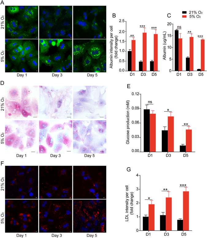
Figure 2.

Physiological oxygen level preserves primary hepatocyte functions. (A) Immunofluorescence staining of albumin (green) in hepatocytes cultured in 21% or 5% O2. Nuclei were stained with Hoechst. Scale bar: 10 μm. (B) Statistical data of the intensity of albumin staining in (A). Data are shown as fold change relative to the 21% O2 group in day 1. Data are Means ± SEM (six random fields) in a representative experiment. (C) ELISA analysis of the secreted albumin levels in culture media of primary hepatocytes cultured in 21% or 5% O2. Data are Means ± SEM (n = 3). (D) PAS staining of the glycogen storage of primary hepatocytes cultured in 21% or 5% O2. Scale bar: 50 μm. (E) Glucose production of primary hepatocytes cultured in 21% or 5% O2. (F) Representative immunofluorescence images of Dil-Ac-LDL uptake (red) in primary hepatocytes cultured in different conditions. Scale bar: 10 μm. (G) Statistical data of the mean intensity of LDL per cell as shown in (D). Data are shown as fold change relative to the 21% oxygen group in day 1. Data are Means ± SEM (seven random fields) in a representative experiment. *p < 0.05, **p < 0.01, ***p < 0.001.
