
Figure 5.
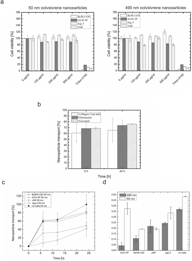
Evaluation of barrier function: Transport of nanoparticles through placental cell barriers. (a) Cytotoxicity of fluorescently-labelled 50 nm and 490 nm polystyrene nanoparticles. (b) Influence of adhesion promoters on the transport of fluorescently-labelled 50 nm polystyrene nanoparticles through 3 µm transwell membranes. Values for transwell inserts without cells were set to 100% of membrane-less wells. (c) Time-trace of transcellular transport after 6 h, 12 h and 24 h of transport of 50 nm fluorescently-labelled polystyrene nanoparticles through BeWo, ACH-3P, Jeg-3 and JAR cell barriers. (d) Apical-to-basal transport ratio after 24 h for 50 nm and 490 nm fluorescently-labelled polystyrene nanoparticles through BeWo, ACH-3P, Jeg-3 and JAR cell barriers. Data points are mean values ± SD for n = 3.