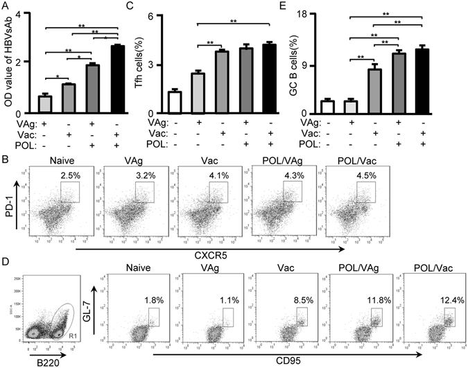
Figure 3.

POL enhanced Tfh cell responses in immunized C57BL/6 mice. (A) 7 days after the 3rd vaccination, the serum of immunized C57BL/6 mice were collected for HBV surface antibody level by using HBV surface antibody kit. (B) 7 days after the 3rd vaccination, the splenocytes were stained with anti-mouse CD4-APC-Cy7/CXCR5-V450/PD-1-PE mAbs. The CD4+ cells were gated for Tfh cells analysis by flow cytometry. The CD4+CXCR5+ PD-1+ cells were counted relatively to total CD4+ cells. (C) The statistical results of Tfh cells were shown. (D) 7 days after the 3rd vaccination, the splenocytes were stained with B220-PE-Cy5/CD95-APC/GL-7-FITC. The B220+ cells were gated for GC B cells analysis by flow cytometry. The B220+CD95+GL-7+ cells were counted relatively to total B220+ cells. (E) The statistical results of GC B cells were shown. Shown in each panel is 1 of at least 3 experiments with similar results. Bar, mean and SD from 3 independent experiments, each using at least three mice per group (n = 3); *p < 0.05; **p < 0.01.
