Figure 1.

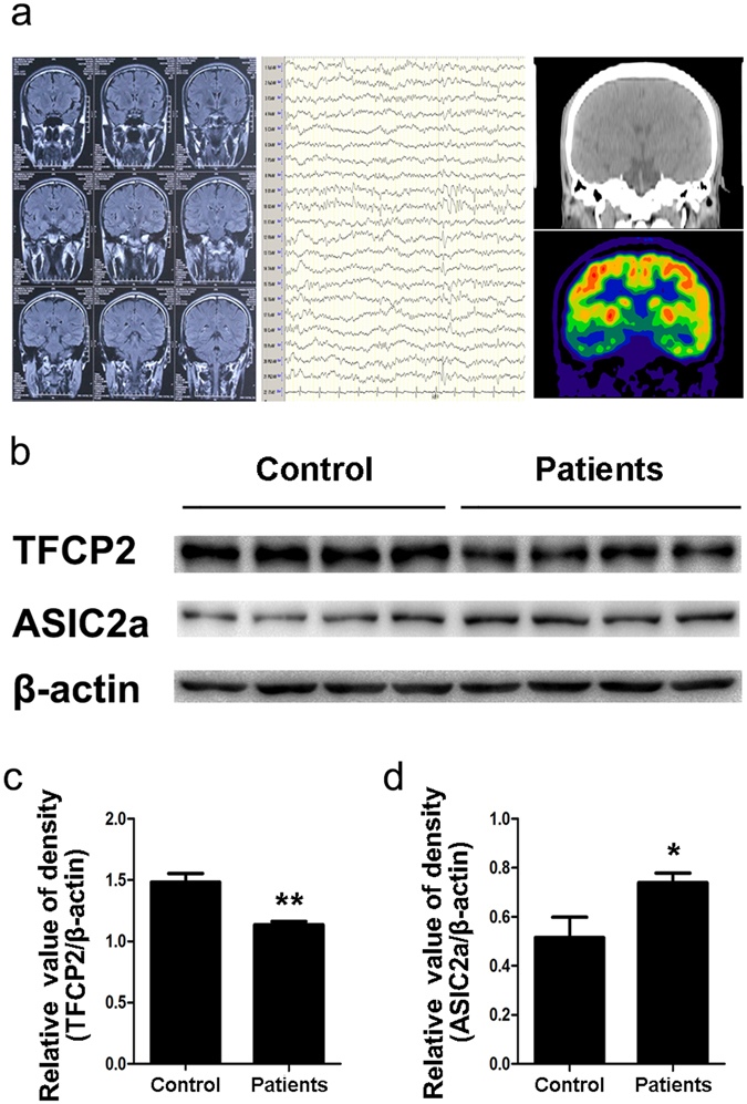
Altered hippocampal TFCP2 and ASIC2a expression with glucose hypometabolism in patients with TLE. (a) Patient 4’s pre-surgical assessment results: magnetic resonance imaging (left) was negative, electroencephalography (middle) showed spike waves in the temporal lobe, and fluorodeoxyglucose positron emission tomography (right) revealed hypometabolic lesions in the right hippocampus. (b) Representative western blot assays of hippocampal TFCP2 and ASIC2a expression in patients with TLE (n = 13) and control patients (n = 10). β-actin was used as a loading control. (c,d) Normalised densitometry bar graphs of TFCP2 and ASIC2a for the control subjects and patients with TLE. The experiments were repeated at least 3 times. Data are presented as means ± standard errors and were analysed using unpaired t-tests. *P < 0.05, **P < 0.01 compared to controls. Uncropped western blot images are shown in Supplementary Fig. 1. Abbreviations, ASIC2a: acid-sensing ion channel 2a; TFCP2: transcription factor CP2; TLE: temporal lobe epilepsy.
