
Figure 2.
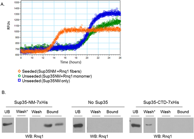
Rnq1 and Sup35 physically interact. (A) ThT kinetic experiments monitor the polymerization of amyloid via enhanced fluorescent emission. Unseeded Sup35 (blue line) polymerizes and aggregates after approximately 17 hours, as does Sup35 incubated with Rnq1 monomer alone (green line). The addition of Rnq1 fibers (orange line) reduces the lag time for Sup35 polymerization to approximately 12.5 hours. Curves represent data from three experiments. (B) Sup35 was immobilized on resin and utilized as bait for a [RNQ +] trap assay. Cell lysates were incubated with the resin, washed with buffer, and eluted. The presence of trapped, untagged Rnq1 was detected via western blot with anti-Rnq1 antibody. Lanes shown are the unbound (UB) fraction, the final wash fraction and the first two elution fractions. “Wash*” indicates an intermediate wash fraction. Blots are representative images from three independent experiments.