
Figure 6.
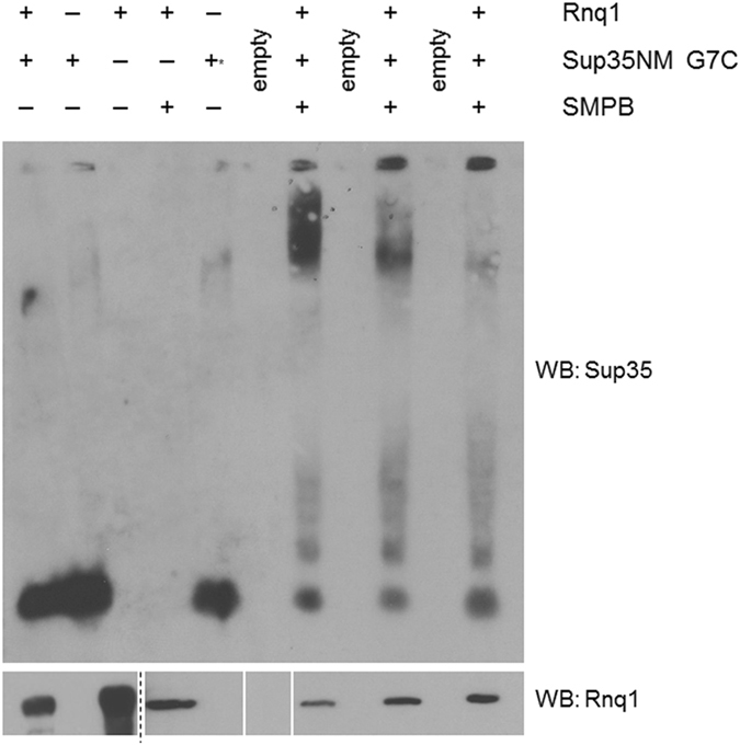
The N terminal region of Sup35 crosslinks to Rnq1. Site-directed crosslinking of Sup35-G7C to Rnq1 created high-molecular weight aggregates as visualized by SDD-AGE. Rnq1 alone, Sup35-G7C alone, and both proteins together without SMPB did not create large aggregates. The “+*” notation in the fifth lane indicates Sup35 monomer following treatment with TCEP. Western blot is a representative image from three independent experiments. In the lower blot, Rnq1 loading was confirmed by SDS-PAGE. Vertical white bars separate non-adjacent lanes of the same blot. The dashed line separates adjacent lanes of the same blot under differing film exposures for image clarity (full blots in Supplementary Figure S4). There is excess Rnq1 in lanes 1 and 3, samples prepared without crosslinker, to adjust for protein that is lost during sample desalting following attachment of the crosslinker (see Methods). Three trials of crosslinking were included in each experiment and the crosslinking experiment was repeated with different batches of purified proteins three times.