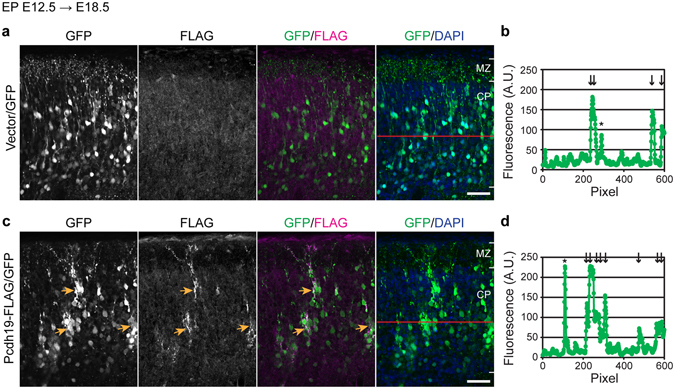
Figure 2.

Overexpression of Pcdh19 induces clustering of neurons in the cerebral cortex. (a–d) pNeuroD-Cre driven expression of GFP without (a,b) or with Pcdh19b-FLAG (c,d) in cortical neurons. DNA transfer was performed by in utero electroporation at E12.5. (a) GFP-labeled neurons were distributed in the cortical plate. (b) Fluorescence intensities along the red line indicated in the rightmost image in a. Arrows indicate GFP-positive cell bodies, and asterisk indicates a neuronal process along the line. (c) GFP-labeled neurons expressing Pcdh19-FLAG formed clusters in the cortical plate. (d) Fluorescence intensities along the red line indicated in the rightmost image in (c). Arrows indicate the GFP-positive cell bodies, and asterisk indicates a neuronal process along the line. Scale bar, 50 μm.
