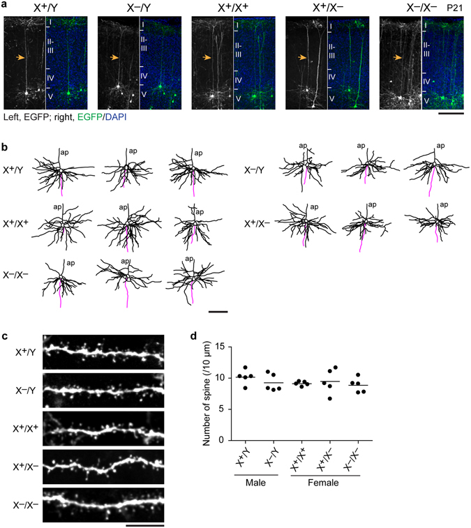
Figure 4.

Formation of apical and basal dendrites and spines of layer-Va neurons in Pcdh19 mutants. (a) Representative layer Va neurons labeled by in utero electroporation of GFP. Arrows indicate the apical dendrite of each neuron. Brains were fixed at P21. (b) Examples of dendritic branches of layer Va neurons. Tracings of dendrites and axons were shown in black and magenta, respectively. ap, apical dendrite. (c) Examples of spines along basal dendrites of layer Va neurons. (d) Number of spines per 10 μm along basal dendrites did not significantly differ among wild-type and Pcdh19 mutant mice (P = 0.6184, n = 5 neurons). Scale bars, 100 μm in (a,b) 10 μm in (c). 100 μm-thick slices were used for analysis.
