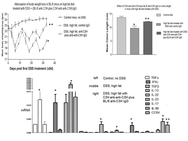
Figure 6.

Inflammatory colitis in mice receiving C3H anti-anti-C3H antibodies plus BL/6 anti-C3H IgG antibodies. The upper left panel shows attenuation of weight loss in C57BL/6mice receiving antibody treatment. * P<.05 compared with control diet; ** P<.05 compared with high fat diet and control Ig (Mann-Whitney U test). The upper right panel shows diminished changes in colon length caused by DSS and high fat diet in mice receiving antibody treatment. * P<.05 compared with control diet; ** P<.05 compared with DSS/high fat diet and control Ig (Man Whitney U test). Finally, data in the lower panel shows attenuation of cytokine mRNA expression in DSS colitis after antibody infusion. Mice received 10µg of each of the antibodies intravenously on days -14 and -7 prior to the commencement of DSS and high fat diet on day 0. * P<.05 compared with control diet; ** P<.05 compared with high fat diet and control Ig (Mann-Whitney U test).
