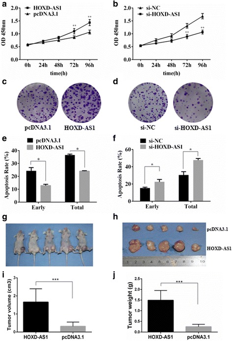
Fig. 3.

The effect of HOXD-AS1 on cancer cell growth in vitro and in vivo. a and b MTT assays were performed on HCCLM3 cells with HOXD-AS1 overexpressed or knocked down. c and d colony formation assays were performed on HCCLM3 cells with HOXD-AS1 overexpressed or knocked down. e and f HCCLM3 cells were transfected with the HOXD-AS1-pcDNA3.1 overexpression plasmid or siRNAs, and apoptosis was induced by the addition of doxorubicin (Dox,1 μM). Flow cytometry was used to determine the apoptotic rates in the different groups. g and h Representative xenograft nude mouse model. HCCLM3 cells were transfected with HOXD-AS1-pcDNA3.1 or pcDNA3.1 control vectors and inoculated into the left or right flank of nude mice. i Tumor volume and j tumor weight were analyzed by the two-sample t test. The data are shown as the mean ± SD, Student’s t test, *p < 0.05, **p < 0.01, ***p < 0.001
