Figure 5.

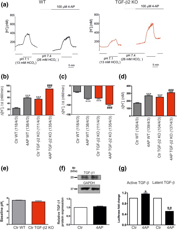
4AP activates extracellular TGF‐βs. (a) Original recordings of intracellular [H+] ([H+]i) in cultured wild type (WT) and Tgf‐β2‐deficient (TGF‐β2‐KO) mouse cortical astrocytes during reduction of external pH and [ ] from 7.4 and 26 mM to 7.1 and 13 mM, respectively, to challenge outwardly directed NBCe1 activity, before and after application of 4AP. Bar plots of the rate of acidification (b), the rate of alkalinisation (c), and the amplitude (d) as measured by changing external pH and [ ] to 7.1 and 13 mM, respectively, and back to pH 7.4 and 26 mM [ ], before and after incubation with 4AP in WT and TGF‐β2‐KO astrocytes (***p < .001, for significant increase, compared to the untreated (Ctr) WT astrocytes, ### p < .001 for significant increase, compared to untreated TGF‐β2‐KO astrocytes, using the Student's t‐test, n = 3). (e) Quantification of baseline pHi in untreated (Ctr) cortical astrocytes derived from either WT or Tgf‐β2 deficient mice (not significant, using Student's t‐test, n = 3). (f) Immunoblotting for endogenous TGF‐β1 protein in control and 4AP‐treated cortical astrocytes (not significant after densitometric analysis of the signal ratio TGF‐β1: GAPDH and Student's t‐test, n = 6). Protein of 50 µg was loaded per lane. (g) The amount of active and latent TGF‐β in the extracellular environment was quantified with the MLEC/PAI‐luciferase assay. Data are given as relative amounts of TGF‐β in the supernatant following 4AP treatment, as compared to the untreated controls (*p < .05 and **p < .01, using the Student's t‐test, n = 3)
