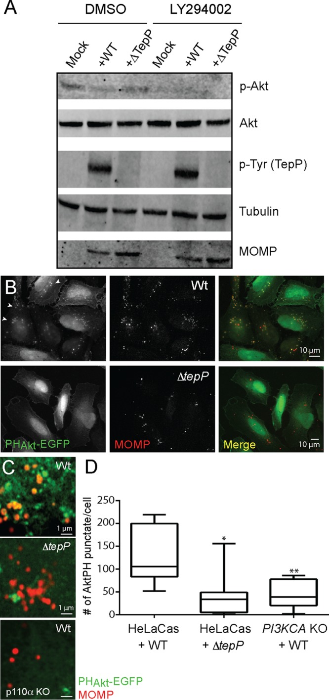
FIG 6 .

TepP activates PI3K at early inclusions. (A) Immunoblot analysis of HeLa-Cas cells infected with CTL2 or ΔtepP::bla C. trachomatis for 4 h indicated that phospho-Akt levels were not significantly altered by TepP. (B to D) Accumulation of PIP3-positive puncta, as assessed by the recruitment of PHAkt-EGFP, in HeLa cells infected with CTL2 but not ΔtepP::bla C. trachomatis. (B) HeLa cells were transfected with PHAkt-EGFP for 24 h, infected with the indicated C. trachomatis strains for 4 h, fixed, and immunostained with anti-MOMP antibodies. (C) Closeup image of clusters of internalized C. trachomatis displaying association with PIP3 (PHAkt-EGFP positive). Levels of bacterium-associated PHAkt-EGFP intracellular puncta were significantly reduced in CTL2-infected PI3KCA KO HeLa cells. (D) Quantification of PIP3-positive puncta in cells infected with CTL2 or ΔtepP::bla C. trachomatis was performed on a per-image basis with 7 to 10 fields in total. Note that punctum formation required the p110α subunit of PI3K. Significance was determined by Student’s t test (*, P < 0.01; **, P < 0.001).
