Figure 6.

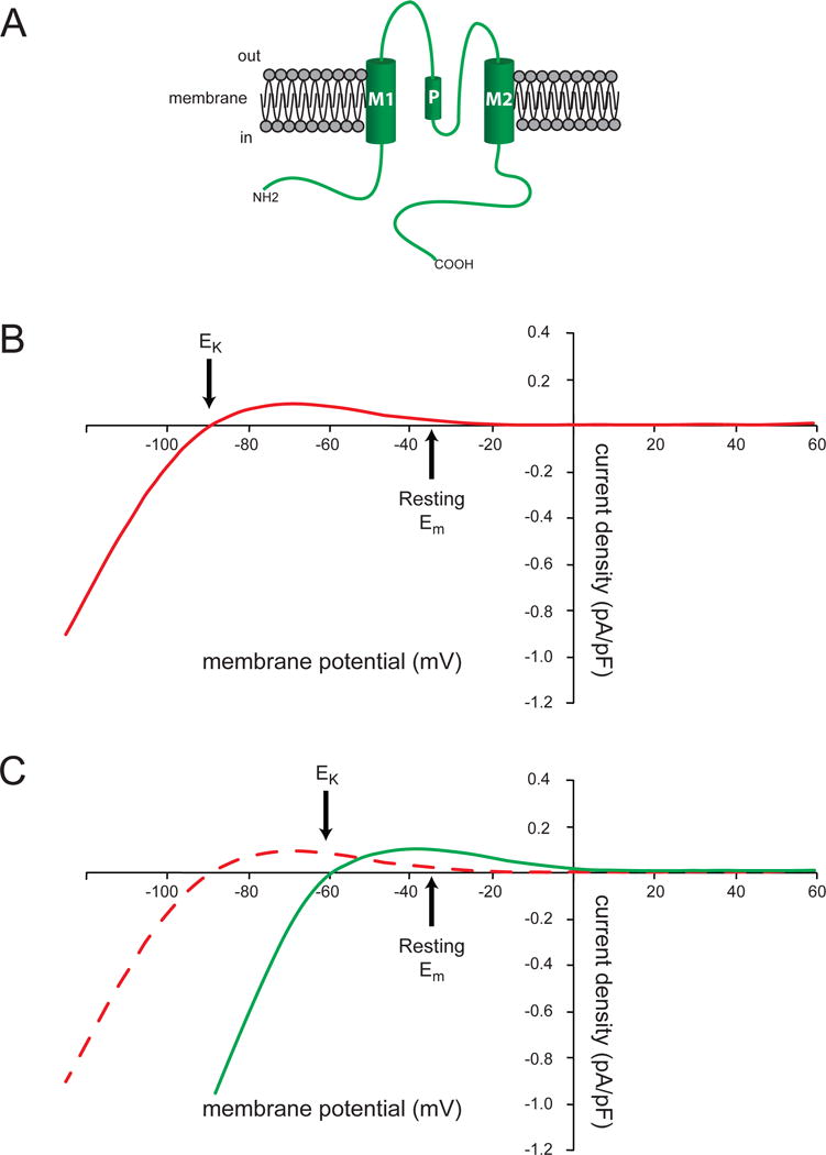
Vascular KIR channels and their currents. Panel A shows the topology of KIR channels with two membrane spanning domains. Functional channels are composed of a tetramer these subunits (see Figure 2). Panel B shows a schematic of the current-voltage-relationship for VSM KIR channels for a cell with 5 mM K+ in the extracellular solution (140 mM K+ intracellular) and is based on data from (Filosa et al., 2006). At membrane potentials more negative than the K+ equilibrium potential (EK, ~−90 mV in 5 mM K+), the channel conducts K+ into the cells, as shown by the negative current density values. At potentials more positive than EK up to ~−30 mV, KIR channels conduct K+ ions out of the cell, and contribute to the resting membrane potential as denoted by the small positive currents at the assumed resting membrane potential of −35 mV. Note that anything that hyperpolarizes the membrane will recruit outward, positive current through KIR channels, effectively amplifying the initial hyperpolarization. Panel C demonstrates the effects of increasing extracellular K+ from 5 mM (red dashed curve) to 15 mM K+ (green solid curve): increased extracellular K+ shifts the EK from −90 mV to ~−60 mV. Note that there is now an elevated outward K+ current at the original resting membrane potential. This enhanced outward K+ current will hyperpolarize the VSM cell membrane from its resting value toward EK, leading to vasodilation.
