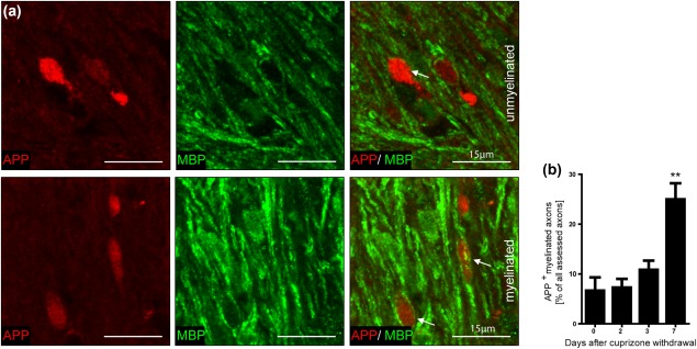
Figure 3.

The percentage of myelinated damaged axons increased during remyelination. (a) Unmyelinated and myelinated APP+ axons were observed in the corpus callosum of mice after demyelination and during remyelination. A representative image of the corpus callosum 3 and 4 days after withdrawal of cuprizone is shown. (b) APP+ axons were analyzed in 30 randomly selected areas with at least one APP+ axon (approx. 50 axons) in the corpus callosum of mice by confocal microscopy. After 1 week of recovery the number of myelinated APP+ axons increased significantly indicating remyelination of damaged axons (**p < 0.01; n = 3‐7; mean ± SEM)
