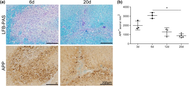
Figure 4.

Significant reduction of acute axonal damage during lesion repair in lysolecithin‐induced demyelination.
(a) Representative images of lysolecithin‐induced lesions in the rat corpus callosum on days 6 and 20 postinjection. LFB‐PAS histochemistry revealed almost complete demyelination after 6 days and almost complete remyelination after 20 days.
APP+ axons were observed in lysolecithin‐induced lesions. (b) The number of APP+ axons in the lesions increased until day 6 and showed a significant decrease on day 20 after lysolecithin injection (*p<0.05; n = 3‐5; mean ± SD)
